Bilix, recognized as a top Innovator’s Pitch Challenge winner at RESI Boston this past September, is making waves in the biotech space with its innovative multi-modality approach to inflammatory and autoimmune diseases. In this interview, Myung Kim, Founder and CEO, shares how participating in RESI Boston helped the company connect with key investors, refine its strategy, and advance its clinical milestones.
Hear firsthand how Bilix is driving progress in complex disease treatment and discover how your company can join the next generation of innovators pitching at RESI London and RESI JPM. Applications are now open.
Commercial interest in targeted epigenetic therapies — agents that target specific genes without altering bases in their sequence or causing double-strand breaks or even single nicks in the DNA — continues to grow, as underscored by the latest financing announced by Epigenic Therapies. The unique selectivity and specificity of targeted epigenetic therapeutics offers compelling advantages over small-molecule epigenetic drugs, which target a specific epigenetic reader, writer or eraser, but affect genes across the genome and affect many diverse tissues, leading to narrow therapeutic windows that make them difficult to develop for conditions outside of cancer.
All of these therapies are designed around an alluring set of simple principles: take a gene-specific DNA-binding domain — zinc-finger proteins (ZFPs), ‘dead’ Cas9 (dCas9) with mutations in its RuvC and HNH endonuclease domains, or transcription activator-like effectors (TALEs) — and tether it via an amino acid linker to an enzymatic effector module. This effector is either an enzyme that directly places or removes a specific epigenetic modification (e.g., TET, histone demethylases or the histone acetyltransferase p300) or a transcriptional activator (e.g., VP16) or repressor (e.g., KRAB).
Epigenetic editing approaches have recently focused on dead CRISPR (dCRISPR) domains fused to various epigenetic effectors, but transcription activator-like effectors (TALEs) and zinc finger proteins (ZFPs) also continue to be explored. Source: TINS
A particularly compelling application for such treatments is genetic disorders of haploinsufficiency (like Dravet’s) or imprinting disorders (like Angelman’s or Prader Willi). There are also many of these diseases where the therapeutic genes would be too large (>4.0 kb) for a traditional AAV gene-therapy approach; in contrast, epigenetic editing machinery can be packaged into an AAV vector.
In a first paper published in Nature, the groups of Kevin Bender and Nadav Ahituv at UCSF (scientific co-founders of Regel Therapeutics) sought to test a targeted epigenetic therapy in patients with SCN2A mutations that exhibit decreased NaV1.2 function. These individuals have impaired action potentials, synaptic transmission and manifest diverse neurological symptoms and seizures, with few therapeutic options, beyond symptomatic anti-seizure medications that have a dizzying range of debilitating side effects.
The UCSF teams leveraged conditional genetic knock-in technolgoy or CRISPRa technology — an AAV-delivered SCN2A-promoter-targeting dCas9 fused to a VP16 activator domain — to upregulate transcription of the SCN2A gene. Using either approach, they were able to boost transcript levels from the healthy SCN2A allele, ameliorating electrophsiological deficits and chemical-induced seizure activity in Scn2a+/− mouse models. Importantly, these effects were seen in adolescent mice, which conventionally have been thought to be too old to respond to treatment. This suggests that rescue of normal dendritic excitability with epigenetic agents at later stages of life might be capable of restoring neuronal function, with implications for patients.
In a separate set of experiments, the authors showed that their epigenetic approach was able to rescue neurophysiological activity in haploinsufficient neuron-like cells from SCN2A-knockout human embryonic stem cells. This cross-species reproducibility provides further confidence that CRISPRa-mediated upregulation could be translated into human treatments.
In a second paper in Nature Biotechnology, a team from Epigenic Therapeutics (Shanghai, China) describes the design and validation of optimized epigenetic regulators (EpiRegs) to silence genes in a precise, durable way without altering genomic DNA. Epigen’s Shaoshai Mao and his collaborators at the Chinese Academy of Sciences and the First Affiliated Hospital of Anhui Medical University tested combinations of TALE- and dCas9-based systems, systematically optimizing effector domains and fusion architectures, looking for effective regulators of gene expression. The best-performing variant, EpiReg-T (a TALE-based system, which eliminates the need for a guide RNA), achieved 98% silencing of target genes in mice, substantially outperforming dCas9-based versions.
Using lipid nanoparticles (LNPs) for delivery, a single administration of EpiReg-T in macaques induced long-term repression of the PCSK9 gene, which encodes a validated target for the treatment of hypercholesterolemia. EpiReg-T reduced PCSK9 expression by >90% and LDL-cholesterol by about 60%, with effects persisting for nearly a year (343 days).
Mechanistically, the team used whole-genome bisulfite sequencing and cleavage under targets and tagmentation (CUT&Tag) to show that EpiReg-T induced stable DNA methylation and repressive histone marks at the PCSK9 promoter. The silencing persisted even after liver regeneration and could be reversed by targeted epigenetic activation. Multiomic analysis in mice, macaques and human hepatocytes confirmed high specificity of the manipulation and minimal off-target effects. Overall, these finding, as well as similar results reported in April by Chroma Medicine, establish epigenetic editing as a promising therapeutic platform for durable and reversible gene silencing.
Overall, targeted epigenetic therapies offer clear safety advantages over small molecules that indiscriminately target all genes under the control of an epigenetic eraser or writer enzymes. They avoid the potential risks associated with creating single- or double-strand DNA breaks associated with CRISPR/Cas9 gene, base or prime editing therapies. And they avoid the insertional mutagenesis risks associated with traditional viral gene therapies. What’s more, in applications requiring gene upregulation in haploinsufficient disease, these approaches maintain the endogenous regulatory context of the functional allele. This is in stark contrast to traditional gene-therapy replacement approaches, where overexpression of an introduced therapeutic gene can often lead to toxicities and immunogenecity.
Of course, questions still linger around the persistence of the changes elicited by these epigenetic agents. Will they persist in patients for long periods — for years or even decades? If they can, then epigenetic therapy may offer compliance advantages over small molecules, antibodies, ASOs or even siRNAs, which have treatment durations of six months or less.
Like all genetic medicines, though, delivery remains the key headache. Thus far, AAV vectors, lipid nanoparticles or ribonucleoproteins (RNP) have all been explored to deliver epigenetic therapies (with some evidence that RNPs might have advantages because they can result in higher dCas9 dosages within target cells). For AAV vectors, the fact that targeted epigenetic therapy might only need to be given once might be an advantage in terms of immunogenicity/neutralization concerns against the vector.
A broader point is that the safety profile of targeted epigenetic editors may offer advantages if AAV vectors are used as delivery vehicles: if the epigenetic agents themselves can be delivered at high dosage (given their intrinsic favorable safety profile and presumed maximal tolerated dose), perhaps AAV vector dosages could be lower than current practice. With many current gene therapies requiring dosages of 1013 or more viral particles/kg in patients, it is increasingly becoming clear that unacceptable liver toxicities arise from the virus at these levels in clinical studies. It will be interesting to follow this space as more agents enter human testing.
By Claire Jeong, Chief Conference Officer, Vice President of Investor Research, Asia BD, LSN
Life Science Nation (LSN) is excited to announce the lineup of investor panels at RESI JPM 2026, taking place January 12–13. These panels bring together leading venture capitalists, corporate investors, and strategic partners to discuss the latest trends, challenges, and opportunities in life science investing. From early-stage “first check” VCs to specialized sectors like Cell & Gene Therapy, AI in Healthcare, and Longevity, RESI JPM offers unparalleled insight into the funding landscape.
Day 1 – January 12
9:00 AM | “First Check” VCs Panel: Learn how early-stage investors evaluate founders, milestones, and the critical first institutional check.
10:00 AM | Medtech Strategics Panel: Explore how strategic investors from leading medtech companies drive growth through partnerships and acquisitions.
11:00 AM | Asia Cross Border Investments Panel: Sponsored by Enterprise Singapore, a RESI JPM Title Sponsor
1:00 PM | Family Offices Panel: Hear how family offices provide patient, mission-driven capital to early- and growth-stage companies.
2:00 PM | Cell & Gene Therapy Panel: Discover where capital is flowing in this transformative area of medicine.
3:00 PM | When Should Companies Exit Stealth Mode?: Gain insight into timing the transition from quiet development to public visibility.
4:00 PM | Aging and Longevity Panel: Learn how investors are deploying capital to solutions addressing the world’s aging population.
Day 2 – January 13
9:00 AM | Partnering with Global Pharma Panel: Understand how big pharma approaches early-stage partnerships and collaboration.
10:00 AM | Women’s Health Panel: Explore investment opportunities in an underserved market with enormous potential.
11:00 AM | AI in Healthcare: Discover how AI is reshaping drug development and care delivery.
1:00 PM | Orphan & Rare Diseases Panel: Learn about unique challenges and opportunities in niche, high-need markets.
2:00 PM | Corporate VC Panel: Hear how corporate venture capital balances strategic and financial goals to accelerate innovation.
3:00 PM | Techbio & Synthetic Biology Panel: Explore investment trends at the intersection of biology and technology.
4:00 PM | 2026 Outlook: VC Perspectives Panel: Gain insight into where investors see healthcare innovation heading in the coming year.
These panels are designed not only to share insights but also to foster dialogue between investors and innovators. If you are interested in speaking on a RESI JPM investor panel, please reach out here.
Registration for RESI JPM 2026 is now open, with Early Bird rates available until October 24. Don’t miss your chance to connect with top investors, strategic partners, and fellow innovators at one of the premier life science partnering events. Register for RESI JPM 2026
In this interview, Caitlin Dolegowski speaks with Cuong Do, Founder and Chairman of M6P Therapeutics, about the company’s groundbreaking lysosomal targeting platform, its applications in rare disease and oncology, and the experience of pitching at RESI Boston.
Caitlin Dolegowski (CD): M6P Therapeutics has achieved what was long thought impossible, delivering proteins to lysosomes. Can you explain the significance of this breakthrough?
Cuong Do (DO): An enzyme called GlcNac-1-phosphotransferase (PTase) is responsible for adding mannose 6-phosphate to the surface of lysosomal enzymes. People have tried and failed for decades to increase the expression of M6P, and everybody gave up. Our co-founder Stuart Kornfeld never gave up. He and his post-doc were able to engineer a variant of PTase that turned out to be 20X more effective than PTase itself in adding M6P to lysosomal enzymes. We built upon this breakthrough to create a platform that is able to create enzyme replacement therapies that have very high M6P content. Furthermore, our gene therapies are the only ones that result in M6P-containing enzymes being produced by the transduced cells.
We expanded upon the innovation and created chimeric antibodies that contain M6P as well. This allows these antibodies (after they bind to the targeted antigens) to be brought to lysosomes in virtually all cells in our bodies for degradation. This is a significant advantage over traditional antibodies relying on Fc clearance by only select immune cells.
CD: You have multiple rare pediatric drug designations and two programs nearing the clinic. What are the most exciting upcoming milestones for your pipeline?
DO: We are preparing to start an Investigator Initiated Trial in Australia for our M021 ERT for Pompe Disease in hopes of obtaining early human data demonstrating M021’s superiority over the standard of care.
CD: How does your lysosomal targeting platform extend beyond rare diseases, particularly in oncology with your chimeric PD-L1 and PD-1 antibodies?
DO: We figured out a way to add M6P to any protein, including antibodies. Our chimeric antibodies can be cleared by virtually all cells in the body since virtually all cells have receptors for M6P. This is especially effective for clearing surface antigens from cell surfaces. Our chimeric PD-L1 antibody is able to clear virtually all PD-L1 from the surface of tumor cells and thus activate T-cells and drive T-cell mediated tumor killing. Our chimeric version of Keytruda is able to remove PD-1 from the surface of T-cells and has shown to be more effective in inhibiting tumor growth in vivo than Keytruda itself.
CD: Can you walk us through your IP position and how it supports your growth strategy?
DO: We have invested heavily in IP that has created a portfolio of 9 patent families, 9 issued patents, and ~20 still in prosecution.
CD: Where are you in your fundraising journey, and what types of investors or partners are you looking to engage with?
DO: We have raised ~$40 million in our Seed and A rounds, which we invested to get our programs to where they are today. We are trying to raise a $5 million bridge now in anticipation of a $50+ million Series B next year. In addition to investors, we want to engage with potential partners who might be interested in our molecules.
CD: How did participating in the Innovator’s Pitch Challenge at RESI Boston help advance your business development or investor connections?
DO: We met a few companies who might be interested in partnering on some of our molecules. We’re continuing the conversations.
IPC Applications are now open for the next Innovator’s Pitch Challenge at RESI London 2025 and RESI JPM 2026, with spots filled on a rolling basis.
By Claire Jeong, Chief Conference Officer, Vice President of Investor Research, Asia BD, LSN
At RESI Boston September 2025, over 50 innovative companies participated in the Innovator’s Pitch Challenge (IPC), providing early-stage life science and healthcare companies with a unique platform to present their technologies and connect with potential investors.
The IPC finalists delivered 6-minute pitches followed by a 7-minute interactive Q&A session with a panel of investor judges. Each participating company also showcased its technology at a personalized table space in the RESI Exhibition Hall, creating additional opportunities for engagement.
A distinctive feature of the Innovator’s Pitch Challenge is its voting system. Registered RESI attendees—including startup executives, early-stage investors, and industry experts— “invested” in their favorite IPC companies using RESI cash provided at check-in. Attendees base their decisions on both the pitch presentations, the companies’ responses during Q&A, meeting with them in the exhibition hall at their table space, and during partnering or ad-hoc meetings at RESI.
Applications are now open for the next Innovator’s Pitch Challenge at RESI London 2025 and RESI JPM 2026, with spots filled on a rolling basis.
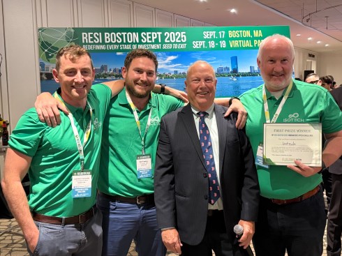
IsoTech Ltd is developing a novel wearable therapeutic medical device that stabilises blood pressure on standing to help older adults avoid dizziness and falls from orthostatic hypotension. The device guides a 30–60-second, on-demand isometric muscle contraction via a discreet retractable tether with visual and haptic cues, turning an evidence-based counter-manoeuvre into a simple, repeatable action without drug side effects. A completed proof-of-concept clinical study demonstrated clinically meaningful blood-pressure stabilisation in a substantial subset of patients with high usability and no unwanted adverse effects. The next phase is straightforward but execution-critical: design-for-manufacture and verification, Class I UKCA/CE technical documentation and light-touch QMS, tooling and pilot build, and a KOL-led seeded launch while preparing reimbursement pilots and strategic licensing options. Protected by patents on mechanism and form factor, ISO-101 aims to become the category-defining, real-time therapy for OH, improving independence for ageing populations and reducing falls-related costs for health systems at scale.
Dr Lochlainn Connolly, Founder – EVP Medical Affairs, Isotech | Dr Neil Fawkes, Founder / EVP R&D, Isotech | Dennis Ford, Founder and CEO, Life Science Nation | Iggy McGowan, Founder / CEO, Isotech
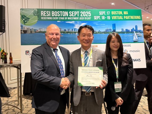
Bilix, founded in Oct 2018, raised $21 million in investments and received $3 million in government grants to develop a new drug for various inflammatory and autoimmune diseases. Our first target is ischemia-reperfusion injury, where unmet medical needs are high and no drugs. Global companies (Novartis, Roche, AstraZeneca, Argenx) are in Phase 2 or 3 trials with single-modality drugs (likely to fail). Our Phase 2a clinical trial (our drug being multi-modality, likely to succeed) runs from 2025.09 to 2026.08. We generate royalty revenue from Classys (a $3B market cap laser medical device company) through exclusive LO in cosmeceutical products. We are currently raising Series B-3 of $6M, of which $ 3 M has been secured. With $25.7 million pre-money valuation, an ROI of >10 times is achievable within three years through IPO/M&A. We can flip to a US company with vetted Series C investments from US/Foreign VCs.
Dennis Ford, Founder and CEO, Life Science Nation | Myung L. Kim, Ph.D., MBA, Founder and Chief Executive Officer, Bilix | Claire Jeong, CCO, VP of Investor Research, Asia BD, Life Science Nation
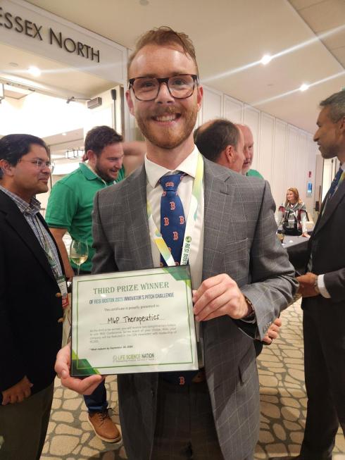
M6P Therapeutics focuses on lysosomes (the natural recycling center for cells) and has the only technology that can deliver any protein to lysosomes – a feat deemed impossible for over 50 years. The company has a deep pipeline, 6 rare pediatric drug designations, and an IP portfolio comprised of 9 patent families, 8 issued patents, and 20 in progress. Our Gaucher Disease ERT is ready for the clinic and our Pompe Disease ERT – which can restore and normalize muscle function – will be ready in 18 months. We also created a platform that can take any antigen-antibody complex to the lysosomes of any cell in the body (instead of relying on Fc clearance by select immune cells) for degradation. Our “chimeric” anti-PD-L1 antibody removes virtually all PD-L1 from the surface of tumor cells while our chimeric version of Keytruda is twice as potent.
Cole Van Vuren, Business Development Manager, Life Science Nation
The participating companies were grouped into 14 sector-focused sessions, with four companies per session. Each session was evaluated by a panel of judges, and the top-scoring companies were recognized for their strong pitches, ability to answer questions effectively, and clear communication of their value propositions.
In healthy epithelial lung cells (top), the CFTR chloride channel translocates from the endoplasmic reticulum to the cell surface, where it participates in the production of mucus to help protect against pathogens. In cells carrying mutant ΔF508 CFTR (bottom), misfolded CFTR cannot reach the membrane and is degraded, resulting in abnormal mucus, microbial infection and lung inflammation. Trikafta restores CFTR function through a dual mechanism: the small molecules elexacaftor and tezacaftor help CFTR fold correctly, whereas ivacaftor improves its gating properties. Source: Lasker Foundation, Michael Welsh
But what about the other 10% of patients who don’t respond to Trikafta, many of whom carry so-called Class I alleles that cannot be rescued by this drug combination? Although a lot of progress has been made, several obstacles lie in the path of effective medicines for people who produce no, or negligible amounts of, CFTR protein.
Class I CFTR alleles. The first three account for ~70% of all patients who don’t respond to Trikafta. Ultra-rare alleles include Y122X, Q1412X, Q493X, and many others.
It should come as no surprise that the main therapeutic strategies for Class I alleles aim to put missing CFTR back into lung cells. Among these strategies, mRNA delivery is the most advanced. VX-522, an RNA therapeutic program from Vertex and Moderna currently in Phase 2, is an inhaled drug that aims to deliver full-length CFTR mRNA to the lung using lipid nanoparticles (LNPs). Two related, competing mRNA delivery programs are at a similar stage of clinical development: ARCT-032 by Arcturus Therapeutics using their LUNAR LNPs; and RCT-2100 by ReCode Therapeutics, which uses a lung-targeted SORT (selective organ-targeting) LNP.
A key feature of RNA-based therapies is that any therapeutic benefit would likely be transient, requiring periodic administration of the medicine to achieve sustained effects. Gene therapy and gene editing have the potential to be a curative, “one and done” procedure. Thus far, however, only gene therapy programs have advanced far enough to be in human testing.
Of these, 4D Molecular Therapeutics’ 4D-710 and Spirovants’ SP-101 use different AAV subtypes designed to optimize delivery to airway basal epithelial cells of a CFTR minigene that lacks the regulatory domain. Both projects are in Phase 1/2 of clinical development.
As the large size (6.2 kb) of the CFTR transgene exceeds the packaging capacity of AAV vectors, Krystal Biotech and Boehringer Ingelheim have launched Phase 1/2 clinical programs using viral vectors with a greater payload capacity: KB407 is a re-dosable herpes simplex virus (HSV)-1 vector with a cargo capacity >30 kb that delivers two copies of the CFTR gene to lung epithelial cells using a nebulizer. BI 3720931 is Boehringer’s inhaled lentiviral vector pseudotyped with Sendai virus F and HN envelope proteins (rSIV.F/HN) engineered to deliver a single copy of the CFTR gene. Further behind in the pipeline, Carbon Biosciences’ CGT-001 is a nebulized non-AAV parvovirus-based vector capable of delivering full-length CFTR gene. Thus far, it has been tested in nonhuman primates and in human bronchial cells in culture.
Companies are also pursuing oligonucleotide therapies to modify disease-causing mutations at the RNA level. SPL84 is an inhaled antisense oligonucleotide (ASO) addressing a splicing defect (cryptic exon; class V mutation) in the ~1,600 CF patients who carry the 3849+10kb C→T mutation. SpliSense has advanced the ASO into phase 2 testing, but it also has in preclinical development an exon-skipping ASO against the class I mutant W1282X. By masking the mutant premature termination codon in exon 23, SP23 induces the splicing machinery to skip exon 23 and stitch together exon 22 and exon 24, forming a partially functional CFTRΔex23 protein.
Gene editing is also beginning to appear on the therapeutic horizon. In July, Prime Medicine announced it had received $25 million in funding to advance prime editors, with a lead program focusing on G542X. Last year, Intellia Therapeutics and ReCode Therapeutics also announced a strategic collaboration to combine the CRISPR pioneer’s Cas9 DNA ‘writing’/insertion technology with Recode’s SORT LNPs. Academic groups have now shown that G542X correction is possible using inhaled LNP- or virus–like particle-delivered adenine base editors. And for RNA editing, at this year’s American Society of Gene & Cell Therapy Wave Life Sciences reported their oligo-based ADAR editors could achieve 21% correction (EC50 = 376nM) of CFTR W1282X nonsense mutations. This is likely a sliver of all the therapeutic activity underway; other programs are targeting mucus itself, which is much thicker than in healthy individuals. If we missed any drug-discovery projects in this space, please let us know!
Despite the plethora of programs, developing genetic therapies against cystic fibrosis patients with class I CFTR mutations faces some stiff translational challenges. For starters, targeted delivery of drugs to lung tissue remains a work in progress. The optimal cell type to be targeted by gene therapy/editing remains an open question, especially as the community continues to identify new cell types in the lung; is it enough to target the more prevalent epithelial cells (alveolar type 2 cells), or will it be necessary to target rarer stem cells (alveolar type 1 cells) to see a long-lasting therapeutic effect? What about the contribution of genetic modifiers and other ion channels known to affect airway dysfunction in CF airway epithelial cells? Also, how to figure out the pharmacokinetics and pharmacodynamics of these disease-modifying therapies in lungs and measure delivery in patients? Specifically, establishing protein expression levels after inhaling a DNA- or RNA-based product would likely require a bronchial biopsy, which is impractical particularly in this fragile patient population.
Last, not unlike most pathologies, new animal and in vitro models with predictive value need to be developed. The use of human bronchial epithelium culture is not as predictive of the efficacy of genetic therapies as it has been for small molecules. At present, the ferret is the gold standard disease model. But it is a time-consuming, challenging animal model, which is only supported by a few groups. All of which slows the path to clinical translation.
Six years after the approval of Trikafta, patient foundations like the CF Foundation, Emily’s Entourage, and the Cystic Fibrosis Trust are devoting increasing resources to translational research to push forward treatments for patients with CFTR Class I mutations who do not respond to potentiators and correctors. The Lasker recognition of the science that led to Trikafta will surely inspire researchers working on those projects to overcome the remaining hurdles.
At RESI Boston, global law firm DLA Piper plays a key role in guiding early-stage innovators through the legal and commercial challenges of scaling in the life sciences. In this interview, Lauren Murdza, Co-Chair of Technology & Life Sciences Licensing & Commercial Transactions, shares why DLA Piper chose to sponsor RESI, what the firm looks for in collaborations, and the trends shaping licensing and commercial transactions today.
Caitlin Dolegowski (CD): What motivated DLA Piper to sponsor RESI Boston, and why do you see value in supporting this conference?
Lauren Murdza (LM): DLA Piper is committed to supporting innovation in the life sciences sector, and RESI Boston offers a unique opportunity to engage directly with early-stage companies and investors. Sponsoring RESI aligns with our mission to be a strategic partner to emerging life science ventures, helping them navigate legal complexities while fostering meaningful connections that drive growth.
CD: From your perspective, what makes RESI a strong platform for connecting with early-stage life science innovators and investors?
LM: RESI creates a unique environment where entrepreneurs, investors, and advisors come together to solve real challenges. For DLA Piper, it’s an opportunity to listen and engage in conversations that matter—how to protect IP, manage data rights, and structure collaborations that attract capital. Those discussions allow us to show how DLA Piper’s integrated approach—combining legal, regulatory, and commercial insight—helps companies accelerate their next milestone.
CD: Can you share what types of companies, technologies, or partners DLA Piper is most interested in engaging with during RESI?
LM: We’re particularly interested in companies developing novel therapeutics, diagnostics, digital health platforms, and medical devices. Our team seeks to engage with founders and executives who are navigating the transition from concept to commercialization and who value strategic legal guidance in areas such as licensing, IP protection, and regulatory compliance.
CD: How does your team at DLA Piper support life science and healthcare companies as they move from early-stage development to commercialization?
LM: DLA Piper supports clients across the full lifecycle of a company—from corporate formation and IP strategy to licensing, financing, and M&A. We help clients identify the core aspects of their technology, assess patentability, and streamline initial filings to create contingent assets that support fundraising. What sets DLA Piper apart is our ability to deliver this seamlessly across jurisdictions, giving clients the confidence that their legal strategy scales with their business.
CD: Are there particular trends or challenges in licensing and commercial transactions that you think entrepreneurs at RESI should be especially mindful of?
LM: We’re seeing three big themes. First, clarity on data and AI rights is critical—investors want to know who owns what and how data can be used, especially across borders. Second, deal structures are evolving, with more options-to-license, milestone-based terms, and royalty monetization to help bridge funding gaps. Finally, regulatory and supply chain issues—from FDA expectations to manufacturing scale-up—are showing up earlier in negotiations. At DLA Piper, we help clients anticipate these challenges so they don’t slow down growth.
CD: What does DLA Piper hope to accomplish through its participation at RESI Boston this year?
LM: We aim to deepen our engagement with the life sciences community, share actionable insights through workshops and panels, and identify promising companies that could benefit from our legal and strategic expertise. RESI Boston is a chance to listen, learn, and contribute to the ecosystem that’s shaping the future of healthcare innovation.
CD: Looking ahead, what excites you most about the current life science innovation landscape, and how does DLA Piper plan to play a role in advancing it?
LM: We’re excited by the convergence of AI, data science, and biotechnology, which is accelerating discovery and personalization in medicine. DLA Piper plans to continue supporting innovators by offering forward-thinking legal solutions and fostering connections that help companies bring transformative technologies to the market.
The firm is focused on therapeutics companies and does not invest in medical devices, diagnostics, or digital health. The firm is open to considering assets of very early stages, even those as early as lead optimization phase. The firm considers various modalities, including antibodies, small molecules, and cell therapy. Currently, the firm is not interested in gene therapy. Indication-wise, the firm is most interested in oncology and autoimmune diseases but has recently looked at fibrotic diseases and certain rare diseases as well.
The firm is opportunistic across all subsectors of healthcare. Within MedTech, the firm is most interested in medical devices, artificial intelligence, robotics, and mobile health. The firm is seeking post-prototype innovations that are FDA cleared or are close to receiving clearance. Within therapeutics, the firm is interested in therapeutics for large disease markets such as oncology, neurology, and metabolic diseases. The firm is open to all modalities with a special interest in immunotherapy and cell therapy.
A strategic investment firm of a large global pharmaceutical makes investments ranging from $5 million to $30 million, acting either as a sole investor or within a syndicate. The firm is open to considering therapeutic opportunities globally, but only if the company is pursuing a market opportunity in the USA and is in dialogue with the US FDA.
The firm is currently looking for new investment opportunities in enterprise software, medical devices, and the healthcare IT space. The firm will invest in 510k devices and healthcare IT companies, and it is very opportunistic in terms of indications. In the past, the firm was active in medical device companies developing dental devices, endovascular innovation devices, and women’s health devices.
A venture capital firm founded in 2005 has multiple offices throughout Asia, New York, and San Diego. The firm has closed its fifth fund in 2017 and is currently raising a sixth fund, which the firm is targeting to be the largest fund to date. The firm continues to actively seek investment opportunities across a […]

 Myung Kim
 Caitlin Dolegowski


 Cuong Do
 Dr Lochlainn Connolly, Founder – EVP Medical Affairs, Isotech | Dr Neil Fawkes, Founder / EVP R&D, Isotech | Dennis Ford, Founder and CEO, Life Science Nation | Iggy McGowan, Founder / CEO, Isotech
 Dennis Ford, Founder and CEO, Life Science Nation | Myung L. Kim, Ph.D., MBA, Founder and Chief Executive Officer, Bilix | Claire Jeong, CCO, VP of Investor Research, Asia BD, Life Science Nation
 Cole Van Vuren, Business Development Manager, Life Science Nation
 Lauren Murdza